Former Department Cognitive Neurology
The former Department of Cognitive Neurology deals with the anatomical and quantifying analysis of the human brain. Research focuses on the functional neuroanatomy of the frontal cortex. Both volunteers and patients are exmamined.
Functional Neuroanatomy of the Frontal Lobe

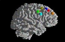
The functional anatomy of the frontal lobes, whose volume amounts to about one third of the human cortex, is not yet fully understood. The involvement of this structure in almost all higher level cognitive processes motivates a research program that aims at a differentiated neuroanatomical description.
Research
Functional Magnetic Resonance Imaging (fMRI)
The most important method is functional Magnetic Resonance Imaging (fMRI), which enables to describe networks of brain regions involved when cognitive tasks are being carried out. Event-related brain potentials (ERPs) provide a closer look at the temporal characteristics of the processes studied. An equally important line of research are patient studies whose goal is to confirm the interpretations of imaging results by providing detailed descriptions of impairments caused by specific lesions of frontal regions. Further, we employ functional Near Infrared Spectroscopy (fNIRS), which is particularly feasible for imaging studies with patients, children and for longitudinal studies. The group's projects target a large number of different aspects of frontal lobe functions, which can be roughly divided into three domains:
- The classic domain of so-called executive functions, including processes such as inhibition, attention, interference or task management, seems to be related to the lateral prefrontal cortex (dual task, imitation, task switching, working memory).
- The most posterior region of the frontal lobe, the lateral and medial premotor cortex, are involved in the organization of movement and its reference to both internal plans as well as external events (action slips, serial prediction and planning).
- Higher level processes, linked to volition and intention, include monitoring, judgment and language. These processes engage regions in the frontomedian wall, an area including, but not restricted to the anterior cingulate cortex (decision under uncertainty, judgement, self performance monitoring, text comprehension).
The overarching aim of the group's projects is to attempt a more fine-grained description of anatomical subdivisions, based on both a differentiated categorization of cognitive functions, as well as a detailed understanding of anatomical constraints.
Clinical Neuropsychology
Clinical studies in the field of neuropsychology and neuro-imaging have been of increasing importance. No matter how well we begin to understand basic and more complex brain functions that allow us to walk, to talk, to think or to memorize, we are challenged and fascinated by an advanced knowledge of the 'pathological state'. The group's projects addressed various aspects of cognitive neurology performed with different techniques, which can be roughly divided into three domains:
- Structural and morphometric characterization of diffuse brain diseases contrasted with focal neurological impairment and its relation to the 'cognitive state'
- Dynamic short-term and long term studies of cognitive dysfunctions associated with several neurological diseases and based on different techniques:
- functional MRI,
- Near-Infrared-Spectroscopy (NIRS)
- Event-related brain potentials
- Reorganization of language function following stroke and in a broader view lateralization of -language function in healthy left- and righthanders and patients with diseases of the CNS
Morphometry and morphological imaging
Morphometric analysis of brain structures aimed to quantify anatomical differences among brains. Macroscopic differences can be expressed in volumetric values for several compartments and substructures such as grey matter, white matter, ventricles and single lobes. In addition, quantitative analysis of the cortical thickness can contribute to a characterization of the normal and diseased brain. The possible applications are various, due to the high number of brain diseases that go together with global or focal tissue loss, such as neurodegenerative diseases, ischemic infarcts, tumors and trauma, for example.
The technique is based on the segmentation of brain structures due to their signal intensity in MR- Imaging. Before the number of voxels of each compartment can be counted, the brain has to be normalized, segmented, and peeled (i.e. cutting off scalp structures) and classes of signal intensity scales have to be defined.
Correlating the morphometrical data with handedness, gender, and language representation as well as with neuropsychological findings in cognitively impaired patients opens a new field of research.
Functional MRI : Reorganization of language function in chronic aphasia
The focus of this research was to determine different mechanisms of language reorganization following aphasia after chronic cerebral stroke. Recovery of language function in patients with aphasia due to infarction in the territory of the left middle cerbral artery has been extensively investigated mainly during the acute stage of the disease. (Weiller, 1995; Heiss, 1999). Functional activation maps -based on PET and fMRI-technique - could visualize different aspects of reorganization. The shift to the subdominant hemisphere and functional recruitment of cortices adjacent to the original language representation area were frequently reported mechanisms of functional recovery in stroke patients with left hemisphere lesions. So far, the relation between imaging data and functional outcome is not fully understood. Moreover, there is an ongoing debate, whether the BOLD signal changes in pathological states reflect neuronal responsiveness. In particular, the influence of perfusion parameter has not been settled yet.
Our research in this field focused on four different aspects of reorganization:
- The size and location of the stroke lesion as well as the infarction type were taken into account and related to the functional activation patterns.
- A comparative assessment of BOLD MR signal changes and MR perfusion imaging was used to determine the influence of regional cerebral perfusion criteria on the BOLD signal change.
- The temporal dynamics of reorganizational changes were investigated in the course of recovery.
- Rare cases of mirrored language organization such as crossed nonaphasia were extensively investigated to understand the underlying physiology of hemispheric dominance.
- The reorganizational changes during language processing following a newly acquired stroke event were compared to activation patterns accompanying a congenital malformation such as Arachnoid cysts in the left temporal fossa.
Near-Infrared-Spectroscopy
Functional near-infrared spectroscopy (fNIRS) allows monitoring of brain activation by measuring changes in the concentration of oxy- and deoxy-haemoglobin as well as changes in the redox state of the cytochrome-c-oxidase by their different spectra in the near-infrared range. Brain activation leads to an increase in cerebral blood flow without a proportionate increase in oxygen consumption, and, consequently, to an increase in the concentration of oxy-haemoglobin and a decrease in the concentration of deoxy-haemoglobin. fNIRS has several advantages in comparison with other imaging methods, such as flexibility, portability, low cost and biochemical specificity. Moreover, patients and children can be repetitively examined.
We used the technique to examine the haemodynamic response in patients with microangiopathy during performance of a Stroop-task and visual stimulation and compared it with the haemodynamic response in healthy controls. Brain activation was investigated with the near-infrared spectrometer NIRO-300 by Hamamatsu. Optodes were placed symmetrically above the dorsolateral prefrontal (Stroop-task) and the visual cortex (visual stimulation). Reaction time was longer in patients with microangiopathy than in age-matched healthy controls during the Stroop-task (p<0.005).
In summary, the haemodynamic response was reduced and delayed in patients with microangiopathy in comparison with age-matched controls during functional stimulation in the lateral prefrontal cortex (Stroop-task) and in the visual cortex. Thus, our data agree with previous reports of altered haemodynamic response in microangiopathy (Terborg 2000, Sabri 1998, 1999).
Event-related brain potentials
Event-related brain potentials (ERPs) are small voltage oscillations recorded by means of electrodes at the scalp, and they reflect electrical activity of the brain time-locked to the ongoing information processing of a particular event, such as the presentation of a stimulus or a (re)action of the subject. ERPs have a dual status allowing to investigate physiological as well as psychological processes, or "they can serve as ... 'windows' on cognition - and they can serve as ... 'windows' on the brain" (Coles, 1989). In most cases, the amplitude of the ERP is very small (microvolts) in relation to the EEG waveform (tens of microvolts). Thus, the event-related EEG activity must be extracted from the background EEG by averaging techniques. In contrast to other physiological measures of brain activity which are based on, e.g., the hemodynamic response of the cerebral vascular system (fMRI, PET), ERPs detect brain activity with virtually unlimited temporal resolution. Furthermore, synchronized activity in properly oriented neuronal populations of only several tens of milliseconds is sufficient to be registered at the scalp. However, the spatial resolution of ERPs is low such that it is not possible to unequivocally determine the neuronal generators of the ERP components on the basis of the scalp-recorded ERPs alone. Therefore, best interpretations can be obtained by combining ERP with hemodynamic neuroimaging methods.
The ERP technique is well-suited for patient studies because it is easy to apply repeatedly such that follow-up studies can be performed. In addition, patient studies with ERPs can reveal information about the function of brain structures, the functional significance of ERP components, and the generators of ERP components.