Nuclear Magnetic Resonance
The NMR Unit is primarily engaged in the development and adaptation of novel magnetic resonance (MR) methods and research into the fundamental biophysics underlying neuroimaging contrast.
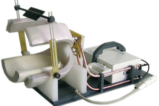
With magnetic resonance (MR) techniques, detailed information is obtained about the anatomy of the brain, its metabolism, or neural activity. Our group is engaged in the development of imaging methods to study these properties as well as of mathematical procedures for image analysis. This also includes hardware components, such as the development of radiofrequency coils for improving the sensitivity in selected MR experiments, such as the investigation of brain tissue specimen. Finally, we conduct research into the biophysics underlying image contrast to gain quantitative data about tissue composition or physiological processes.
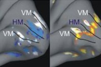
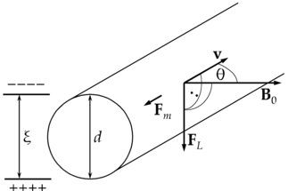
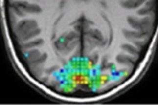
Of ongoing interest to us is the precise imaging of parameters characterizing perfusion, including regional blood flow and blood volume. This permits mapping of activity patterns to investigate laminar-specific information processing in the cortex. Another research area focuses on the microstructure of brain tissue. Axons, which are responsible for signal transmission in the brain, are surrounded by a myelin sheath, which consists of layers of membranes and acts as an electrical insulator. It is of fundamental importance because it drastically increases the speed of transmission. Information on myelin can be obtained indirectly by studying the relaxation of water molecules in close contact with the membranes or by investigating physical tissue properties, such as the magnetic susceptibility.


