Highly detailed 3D brain reconstructions
A comprehensive map of the human brain is a long-held goal of neuroanatomists. Noninvasive imaging techniques such as magnetic resonance imaging (MRI) do allow scientists to study the healthy living human brain. However, they provide only limited anatomical detail. A higher level of detail can be obtained by microscopy on the brains of deceased donors. But that, in turn, usually focuses on small brain structures imaged in 2D. Now, a team of scientists from the University of Amsterdam and the Max Planck Institute for Human Cognitive and Brain Sciences has combined MRI and microscopy. This has produced 3D images of two complete brains with an unprecedented level of detail.
. Since the whole brain is captured, it simplifies the link to in vivo magnetic resonance imaging. It also facilitates the precise localization of small but crucial structures such as the thalamic nuclei.")
Thanks to these images, it is now possible to virtually dissect the brain. "We are excited about all the possibilities this can open up for the field. Instructors can use the datasets for neuroanatomy training or virtual dissection, for example. And the ability to compare MRI results to individual proteins will give researchers more insight into poorly understood MRI observations and provide more anatomical details about small brain structures," said Anneke Alkemade, a neuroscientist at the University of Amsterdam and first author of the underlying publication in the journal Science Advances.
"It allows us to better link the MRI images to the underlying biological microstructures," adds Nikolaus Weiskopf, director at MPI CBS and also one of the study authors. "By improving this link, we will arrive at new imaging biomarkers that can detect pathological changes much earlier than today. Just what it takes for early diagnosis and treatment of neurodegenerative diseases like Parkinson's and Alzheimer's." Co-author Evgeniya Kirilina, also of MPI CBS, adds, "It's a unique resource for studying very small but very important brain nuclei that supply the brain with the neurotransmitter dopamine."
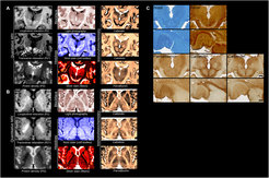
For their work, the researchers used a 7-tesla ultrahigh-field MRI system, which has a stronger magnet than MRI systems routinely used in hospitals. The MRI software was programmed by the researchers specifically for these studies to account for the differences between living and preserved tissue. When the tissue was cut, each section was photographed individually so that the deformation of the tissue could later be digitally corrected in microscopic sections. The individual brain sections were placed on specially ordered glass slides and processed using custom-built laboratory equipment.
After digitizing each slide, researchers created new algorithms to correct the tissue deformations created by the cutting and microscopy. After weeks of uninterrupted calculations, the researchers were finally able to create complete reconstructions of two individual brains.
The researchers have now made the data they obtained available free of charge in the spirit of open science and as a service to the scientific community. Scientists and other interested parties from around the world can now travel through the resulting 3D reconstructions of the human brain.


