Three questions to...
... Laurentius Huber, winner of the Otto Hahn Medal 2016.
Laurentius Huber could enable a more precise look into the brain at work with his dissertation at Max Planck Institute for Human Cognitive and Brain Sciences. Therefore he will be honored as one of thirty young researchers by the Max Planck Society.
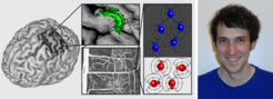
 to minimize alignment with the external magnetic field, while tissue water protons (illustrated in blue) tend to point to a preferred direction.")
Mr. Huber, your PhD thesis recently won the Otto Hahn medal. Could you explain to us what is special about your work?
In my research, I’m trying to develop non-invasive neuro imaging methods that can map brain activity with the highest possible precision. My main focus is on a method which indirectly also measures the brain activity where blood vessels in active brain areas increase their volume to transport more oxygen and glucose, according to demand, as a proxy for neural activity change. Measuring small blood volume changes in the immediate vicinity of neurons makes this method particularly specific to localised neural activity.
A prominent feature of the new method, known as SS-SI-VASO, came from a fortunate coincidence of the synergy effect between the timing of blood flow dynamics and blood magnetization properties (spin lattice relaxation), which allows particularly high spatial resolutions.
More specifically, it takes about 2 seconds for the magnetization from blood water protons to align with the magnetic field of the MR scanner. By lucky coincidence, it also takes around 2 seconds for blood to flow from the neck into the tiny brain capillaries. In the new method, we are able to match the timing of the gradient and radio field switching to match the blood flow and spin dynamics in such a way that we can manipulate the magnetization of blood flow differently as the magnetization of stationary (non-moving) tissue. The timing is adjusted so that a single radio frequency pulse annihilates the net magnetization of moving blood, while the steady-state magnetization of stationary tissue receives many more radio frequency pulses and tends to be more aligned along the magnetic field. This trick can amplify the functional signal of blood volume mapping and improves the signal and the resolution by up to 8 times. With the additional signal available, we can now also make brain activity within two to four millimeters thick cortex visible.
What is your motivation to be engaged in improving MRI methods?
My original motivation to devote myself to brain science comes from the time when I was studying basic physics. I was fascinated to learn that physical methods are needed to study abstract concepts such as cognition and consciousness. The motivation to look into blood volume mapping methods was rather accidental during my Master’s project supervised by Prof. Robert Turner. The motivation to continue working on blood volume mapping methods together with my dissertation advisor, Harald Möller, came from our hope to improve this to the extent that it could overcome specific limitations of conventional neuroimaging methods. Specifically, we wanted to minimize unwanted artifacts from large draining veins in order to improve the spatial specificity of activity mapping at high resolutions.
The award-winning thesis is in the bag. What’s next?
Working in the field of neuroscience is a lot of fun and gives me the chance to continuously learn more about how the brain works. If possible, I would like to stay in the field of Neuroscience long-term. I’ve recently started working at the National Institute of Health in Bethesda, close to Washington D.C., on potential applications of the new imaging method with my new advisor Peter Bandettini. We are using the high resolution of this new method to track the basic functional building blocks of the brain’s cortical layers and columns. In doing so, we hope to investigate directional functional connectivity to see how different brain areas are functionally interconnected.