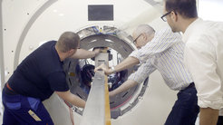
The super brain scanner
It is one of three of its kind worldwide and the only one in continental Europe: The CONNECTOM MRI scanner. Thanks to its unparalleled gradient strength, it will reveal information about the inner wiring of the living human brain that is crucial for the transfer of information in our heads. As of today, the CONNECTOM at Leipzig’s Max Planck Institute for Human Cognitive and Brain Sciences is ready for action.
Siemens MAGNETOM Skyra Connectom A
What does the brain actually look like? Our picture is still coarse when it comes to the routing and the wiring of its nerve fibres. We still know very little about the relation between the exact anatomy and specific functions of the brain, that is, the functional neuroanatomy.
The Max Planck Institute for Human Cognitive and Brain Sciences (MPI CBS) aims to explore these central questions with latest technology: The Connectom MRI scanner—one of the best of its kind worldwide for understanding the inner wiring of the brain and the only one in continental Europe. In fact, one of the leading neuroscientists, Prof. Derek Jones from the Cardiff University, recently called it the Hubble Telescope of Neuroscience. As of today, the long-awaited Connectom at MPI CBS is up and running.
Although it may look like a conventional MRI scanner from the outside, it entails a technology that has a lot to offer: It will live up to its name by providing precise information about the connections in the brain, such as how the fibres run in the depth of our brains, where they cross each other, where they change their direction, where they reach the surface again and enter the cortex, and which brain areas exchange information with each other. “With this technology, we can precisely follow the route of the nerve fibres in the brain that connect single brain areas with each other like data highways”, explains Prof. Nikolaus Weiskopf, director at MPI CBS and scientific expert on the Connectom. “They are the crucial structures for understanding the transfer of information in the brain.”

Its secret lies in its especially high magnetic gradient field. With 300 millitesla per metre, it is almost four times higher than that of the best current clinical MRI scanners. Based on the principle of diffusion-weighted MRI, it is therefore able to non-invasively measure the diffusion of the water molecules in the brain and can delineate fibre bundles much more precisely. To this end, it capitalises on the fact that cell membranes or other barriers can influence the water particles’ movements that can make them move relatively faster parallel to the nerve fibres rather than perpendicular. Derived from diffusion measurements along different directions, neuroscientists can reconstruct the route, shape and size of the nerve fibres – without being able to directly see the axons measuring only a few micrometres. “When it comes to defining the exact course of the nerve fibres, the Connectom is in a class of its own, a milestone in brain sciences and modern medical technology”, says Prof. Harald Möller, the institute’s technical expert on the scanner that was developed by SIEMENS Healthcare.
The Connectom’s capability does not just lie in encoding the tracts of our brain. With a resolution up to ten times higher than that of standard scanners, it will also shed light on unexplored anatomical microstructures of the living human brain. “We want to reach a level of about 0.6 millimetres”, states Prof. Weiskopf. “By doing so we could finally gain a much sharper insight into the cortex which is just three millimetres thick and crucial for our higher cognitive abilities. Maybe we can even reach a level of precision that is high enough to measure interindividual differences.“ Why are some people more fearful than others? Why is one person able to remember things better than another person? Why do some people find it easy to learn a foreign language in no time at all whereas others struggle? These are questions that the Connectom will help us to address in years to come.
Even unresolved issues regarding the adaptability of the brain, known as brain plasticity, could be investigated more directly. For example, which microstructures in the brain change when a person relearns a certain movement after a stroke? “We should bear in mind that each time we succeed in further magnifying these structures, we will master an unprecedented physical-technological scientific challenge”, Prof. Weiskopf adds.
But it is not just the Connectom that will deliver groundbreaking images of the brain. The existing MRI scanners at Leipzig’s MPI CBS will offer entirely new opportunities to combine the special qualities of each machine. “Each scanner is able to reveal different structures and layers. It is therefore our goal to use the images to create a unified biophysical model of the brain structure”, explains the neurophysicist. “A thinning myelin layer of the axons and an increasing iron concentration, for instance, are early signs of ageing. The former can be measured by the help of the Connectom, the latter by the 7 T Magnetom and its seven tesla magnetic field strength."
The combination of these cutting-edge technologies offers neuroscientists at MPI CBS not only the chance to understand in much more detail the early stages of neurodegenerative diseases such as Alzheimer’s disease, but it also opens a new window to fundamental human neuroscience research. “I am convinced, with its help we will deepen our understanding of the human brain by many dimensions and in turn we will abandon many of our previous assumptions. In particular, regarding the so far unknown fibre tracts which have been proven as the underestimated, actually decisive brain structures in recent years”, says Prof. Angela D. Friederici, Vice President of the Max Planck Society and likewise director at MPI CBS in Leipzig as one of the leading locations for neuroscience. “We see ourselves as adhering to our key principle—‘Insight must precede application’.“


