Projects

References


Investigating the aging brain

Before examining pathological processes, one has to understand the normal functioning of the brain. Additionally, one should explore changes during cognitive development and healthy aging. The project addresses these issues by investigating the neural correlates of attention and executive functions during adolescence and aging with optical imaging (functional NIRS) and fMRI. In particular, we apply tests investigating functions of the frontal lobes, for example, the Stroop color word interference and Stop signal task.


Losing your self ...

has been related to the frontal lobes, namely frontomedian regions. This peculiar human ability requires the prediction of mental states and behavior of others, known as “theory of mind” or “mentalizing”. Moreover, self-monitoring, processing/evaluation of internal mental states, perception of pain and emotions, and sustaining personality and self have been associated with frontomedian regions. One dementia syndrome, frontotemporal dementia, is characterized by deep alterations in behavior and personality. We aim to isolate its neural correlates by systematic and quantitative meta-analyses of imaging studies and by investigating MRI and FDG-PET data in our own patient cohort. Indeed, we have been able to demonstrate that frontotemporal dementia specifically affects frontomedian neural networks related to social cognition. Moreover, we dissociated its neural substrates from the substrates of the other two subtypes of frontotemporal lobar degeneration, semantic dementia and progressive non-fluent aphasia, involving mainly language disturbances. Future studies will investigate the neural correlates of executive functions and behavioral deficits in these disorders using MRI and FDG-PET. The project contributes to the placement of the disease in a framework of cognitive neuropsychiatry and may suggest new approaches for therapy. Another frequent disorder is also characterized by alterations of the frontal lobes: Traumatic brain injury. Recently, it was suggested that long term outcomes mainly depend on social cognition. Accordingly, we investigated frontomedian alterations in this disease, namely diffuse axonal injury. We were able to show specific deficits such as inhibition of imitative response tendencies and evaluative judgements in frontomedian tasks using fMRI.


Forgetting your self
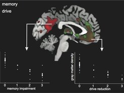
This projects aims to characterize the neural correlates of memory. First of all, Alzheimer’s disease and its prodromal stage mild cognitive impairment (MCI) are the focus of this project. We isolated their neural correlates by means of systematic and quantitative meta-analyses of imaging studies. Furthermore, we identified imaging markers predicting the conversion from MCI to Alzheimer’s disease. Now, we are proving hypotheses in our patient cohort with multimodal imaging (MRI & FDG-PET) and in the comprehensive longitudinal study LIFE at the University of Leipzig. This study explores predictors of conversion from MCI to Alzheimer’s disease in a large cohort of approximately 4,000 subjects during a 2.5 year period. Here, we will focus on the importance of social cognition and emotion comprehension for healthy and pathological aging by combining imaging (MRI, optical imaging) and genetic approaches (polymorphisms). Beside Alzheimer’s disease, heart attacks and resuscitation might lead to severe impairments of memory. Accordingly, another project explores the neural correlates of memory and executive/behavioral deficits in these disorders. Likewise, we are investigating neural networks and their interrelations during unconsciousness due to anesthesia.


Stimulating your self

Parkinson’s disease is a movement disorder that is related to dysfunctions in dopaminergic circuits. Frequently, it leads to cognitive deficits. The project explores effects of dopaminergic agents (L-DOPA) or deep brain stimulation on the blood oxygenation level dependent (BOLD) signal in Parkinson’s disease during a simple finger-tapping paradigm and in task-free situation (in order to investigate low-frequency oscillations). Here, we are cooperating with Robert Jech from the Department of Neurology at the Charles University in Prague.


Investigating the inattentive brain

Optical imaging (NIRS) is a method that is particularly suited to investigations of cognitive development and its diseases. Accordingly, we applied the method to children suffering from attention deficit hyperactivity disorder (ADHD). We were able to demonstrate that functions of the prefrontal cortex are altered during a Stroop task. Subsequent studies aim to identify treatment effects. Another part of this project investigates cerebral microangiopathy that finally leads to vascular dementia. We were able to demonstrate that alterations in frontal cortices are closely related to attentive and executive deficits. Besides optical imaging, we are applying MRI and SPECT.


Investigating the oscillating brain

This project investigates changes of spontaneous hemodynamic oscillations during healthy aging and dementia. Recently, using optical imaging (NIRS), we were able to show that spontaneous oscillations specifically around 0.1 Hz decrease in the visual cortex of the aging brain and in vascular dementia (cerebral microangiopathy). Interestingly, this kind of analysis in the frequency domain was much more sensitive in detecting changes than functional visual stimulation. Hence, we aim to establish our approach as a screening method to investigate vascular reagibility. In the future, we want to extend these approaches to optical topography, covering the whole outer brain & fMRI. Moreover, we will examine patients with Parkinson’s & Alzheimer’s Disease and explore the predictive potential of this method to address changes from mild cognitive impairment (MCI) to Alzheimer’s disease. Finally, we want to examine alterations in transgenic mouse models of healthy aging and Alzheimer’s disease.


Measuring the right signal

Methodological issues are of crucial importance in brain imaging. Accordingly, our group investigates influences of normalization procedures for FDG-PET data on diagnosis and differential diagnosis of dementia. Moreover, we examine the impact of different analysis approaches on magnetic resonance imaging (MRI) data, namely with regard to voxel-based morphometry (VBM).


Enlightening the brain

Optical imaging (NIRS) has been introduced to cognitive neuroscience in recent years. It has decisive advantages in comparison with other imaging methods, namely easy application, insensitivity to movement artifacts and high temporal resolution. Accordingly, it is recommended for studies of cognitive neurodevelopment investigating neonates and children, and for researchers in psychiatry. We have developed various standard analysis approaches for this method, overcoming limitations such as the high variability of the so called “differential pathlength factor” (DPF). We investigated the reliability of the method and applied optical imaging in various studies including psychiatric and neurological patients (see also other projects). Furthermore, we explored the foundations of the neurovascular response (post-stimulus undershoot of the BOLD signal) with a multimodal imaging approach (plus fMRI).


Exploring glia in schizophrenia and depression

In recent years, researchers in the neurosciences have changed their focus of attention from neurons to glia. This “glial turn” has revealed new approaches and hypotheses for the healthy brain and its disorders. Recently, mood disorders, namely major depression, have been described as diseases mainly affecting glia and particularly astrocytes. We have validated the glial hypothesis of mood disorders in humans by measuring serum markers with meta-analyses, and cell culture models. We were able to show that the glial marker protein S100B is elevated in major depression. These elevations seem to be related to the severity of depression and might be used as treatment markers. In contrast, the neuronal marker protein neuron-specific enolase (NSE) is unchanged. However, increases of S100B are not specific for mood disorders. We and others detected the same alterations in schizophrenia that were particularly related to the “deficit syndrome” characterized by “negative symptoms”. Now, cell culture models of astrocytes, which we have also previously used to investigate the blood-brain barrier, are underway exploring drug effects specifically.


The dark side of cognitive neuroscience

Cognitive neuroscience has detected the neural correlates of various cognitive functions such as memory, attention, language, social cognition and executive abilities (“positive phrenology”). In recent years, researchers in cognitive neuroscience have even tried to go beyond these issues by exploring the neural correlates of consciousness and free will. The project discusses these approaches by developing a “negative phrenology”. This research project answers the question of which kinds of cognitive functions cannot be addressed by classical experimental approaches. However, in its second step it tries to go beyond “negative phrenology”. Here, we suggest and investigate data-driven approaches and the incorporation of phenomenological or first-person data into cognitive neuroscience.


Go to Editor View