Projects
Measuring the right signal

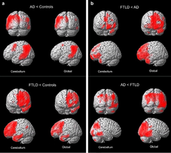
Methodological issues are of crucial importance in brain imaging. Accordingly, our group investigates influences of normalization procedures for FDG-PET data on diagnosis and differential diagnosis of dementia. Moreover, we examine the impact of different analysis approaches on magnetic resonance imaging (MRI) data, namely with regard to voxel-based morphometry (VBM).
Enlightening the brain

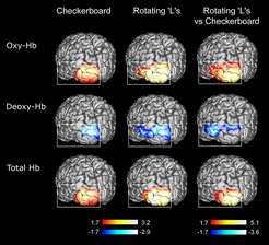
Optical imaging (NIRS) has been introduced to cognitive neuroscience in recent years. It has decisive advantages in comparison with other imaging methods, namely easy application, insensitivity to movement artifacts and high temporal resolution. Accordingly, it is recommended for studies of cognitive neurodevelopment investigating neonates and children, and for researchers in psychiatry. We have developed various standard analysis approaches for this method, overcoming limitations such as the high variability of the so called “differential pathlength factor” (DPF). We investigated the reliability of the method and applied optical imaging in various studies including psychiatric and neurological patients (see also other projects). Furthermore, we explored the foundations of the neurovascular response (post-stimulus undershoot of the BOLD signal) with a multimodal imaging approach (plus fMRI).
Exploring glia in schizophrenia and depression

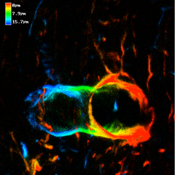
In recent years, researchers in the neurosciences have changed their focus of attention from neurons to glia. This “glial turn” has revealed new approaches and hypotheses for the healthy brain and its disorders. Recently, mood disorders, namely major depression, have been described as diseases mainly affecting glia and particularly astrocytes. We have validated the glial hypothesis of mood disorders in humans by measuring serum markers with meta-analyses, and cell culture models. We were able to show that the glial marker protein S100B is elevated in major depression. These elevations seem to be related to the severity of depression and might be used as treatment markers. In contrast, the neuronal marker protein neuron-specific enolase (NSE) is unchanged. However, increases of S100B are not specific for mood disorders. We and others detected the same alterations in schizophrenia that were particularly related to the “deficit syndrome” characterized by “negative symptoms”. Now, cell culture models of astrocytes, which we have also previously used to investigate the blood-brain barrier, are underway exploring drug effects specifically.
The dark side of cognitive neuroscience

Cognitive neuroscience has detected the neural correlates of various cognitive functions such as memory, attention, language, social cognition and executive abilities (“positive phrenology”). In recent years, researchers in cognitive neuroscience have even tried to go beyond these issues by exploring the neural correlates of consciousness and free will. The project discusses these approaches by developing a “negative phrenology”. This research project answers the question of which kinds of cognitive functions cannot be addressed by classical experimental approaches. However, in its second step it tries to go beyond “negative phrenology”. Here, we suggest and investigate data-driven approaches and the incorporation of phenomenological or first-person data into cognitive neuroscience.