Functional brain mapping by metabolic demand

MRI methods can now provide reasonably accurate maps of cerebral blood flow changes due to brain activity and respiratory challenges. The blood oxygenation level dependence (BOLD) contrast has already proven its utility in functional brain imaging and found widespread use. Very high field strengths benefit BOLD imaging through increased contrast, improved localization to the activation site and possibility of high resolution acquisitions. Using a combination of these techniques, maps of brain oxygen consumption can be derived, providing a uniquely direct quantification of how much work the brain is doing. Only at very high field strength is this technique sensitive enough to be useful in cognitive science. We will then be able to decide unambiguously whether, for example, the brain's activity is actively inhibited in some local region, rather than merely experiencing a local decrease in blood flow that increases blood deoxygenation. The technique will allow the investigation of the changes in brain function associated with learning or the progression of certain diseases.
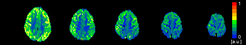
Figure shows MRI maps of cerebral blood flow acquired at 7 T in a normal human brain.
Internal Collaborators include:
Prof Dr Harald Moeller
Dr Derek V.M. Ott
External Collaborators include:
PD Dr Wolfgang Heinke - University of Leipzig Medical Faculty, Leipzig, Germany
PD Dr. Matthias Günther - mediri GmbH, Heidelberg, Germany
Dr Jan Warnking - Institute of Neurosciences, Université Joseph Fourier, Grenoble, France
Dr Kamil Uludag - Max Planck Institute for Biological Cybernetics, Tübingen, Germany
Dr David L. Thomas - University College London School of Life and Medical Sciences, London, UK