Analysis and Interpretation of MRI data
Analysis and interpretation of structural MRI data

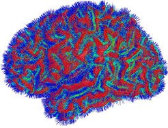
Image analysis is a core technique for extracting information about brain structure from MRI data. To obtain a better understanding of cortical fine structure we need to digitally separate the cortex from other brain tissue such as white matter, non-cortical grey matter, dura, vasculature or cerebral spinal fuild (CSF). Once the cortex is segmented and represented in a surface-based model, a primary goal is to subdivide it into different areas based on very high-resolution MRI data acquired on our 7T scanner with various image contrasts.
Analysis and interpretation of functional MRI data
A principal goal in functional magnetic resonance imaging (fMRI) is the detection of activation areas in the human brain. In a typical fMRI experiment, human volunteers are subjected to experimental stimuli and are asked to respond to them while several hundred images are recorded by an MRI scanner at a rate of about 1 to 2 seconds per image. These image sequences are then analysed using standard statistical techniques to reveal areas in the brain that are significantly activated when a stimulus condition is contrasted against some baseline condition. The result of such an analysis is an activation map that shows the degree of statistical significance with which each pixel can be considered to be activated.
Activations maps of this type are routinely generated in our institute as well as in many other institutions around the world. Even though they are of large value for neuroscience they leave many questions unanswered. For instance, such maps show us which brain areas are involved in a cognitive task but they do not reveal interdependencies between these areas.
We plan to develop new methods for just this purpose. Since current methods explain only about 10 percent of the overall signal variance there is a very good chance that the information we seek is buried somewhere in the remaining 90 percent.
Understanding sulcal patterns
Another research aim is to further our understanding of human sulcal patterning. The folding pattern of the cerebral cortex and its relation to functional areas is notoriously variable. It would greatly assist the mapping of structure to function to identify 3D topographical cortical features that bare a consistent relationship to functional areas. Based on our previous work, we hypothesise that the deepest parts of the sulcal fundi might be the most likely to satisfy these requirements. One of our research aims is to investigate this hypothesis.
Combining information from different sources
A prerequisite for all of the above research is a high-precision geometric alignment between data sets of different types. Methods for image alignment are currently being evaluated.

External Collaborators include:
Alan Colchester, University of Kent

