High resolution mapping of brain white matter
A technique originally developed by Professor Robert Turner, single-shot diffusion-weighted MRI, has turned out to provide very useful information regarding brain connectivity. Cortical areas, and deep brain structures, are joined together by long myelinated nerve fibres called axons. Because water molecules can move more easily along these fibres than across them, measurements of water diffusion in the brain using MRI can show the orientation of axonal fibre bundles.
We also explore diffusion in grey matter. We have extracted the main diffusion direction from DTI data at 3T. This has helped to segment the amygdala, a large region of apparently undifferentiated white matter. Using MRI at 3 T and 7 T we are characterizing nerve fibre directions in the brain with millimeter precision, and we are beginning to correlate their terminations with the results of functional mapping. In principle the higher sensitivity available at 7T will give us improved results, but further improvement in imaging sequence design and image processing are vital.

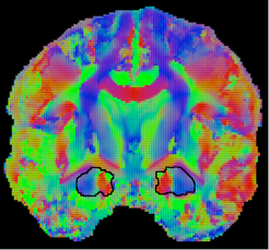
Diffusion in brain tissue is more complicated than in pure liquids, having at least two components. In an ongoing collaborative project with the University of Leipzig, we are trying to understand more about the physical basis of this phenomenon. We use the powerful magnetic field gradients (up to 35T/m) available with the University’s NMR Spectrometer, together with MRI techniques and novel analysis methods. Figure shows a coronal section from a single subject. Vectors represent the main direction. The colour coding represents the mean fibre direction in each image pixel.