WP4 UPÉCS: Clinical translation
Background
Prof. Norbert Kovács leads the Movement Disorder Unit of the University of Pécs (MDU Pécs), which is one of the largest tertiary care centres in Hungary for PD and other movement disorders. MDU Pécs not only provides high quality clinical activity and integrated care for its patients, but also has a wealth of experience in clinical neuroscience research. Dedicated 3T MRI for research, a full neurophysiology and sleep diagnostic laboratory, and full-time neuropsychologists and Parkinson's nurses. Since 2013, MDU Pécs runs a prospective database of over 2,000 PD patients, which complies with the relevant ethical and data protection guidelines. Subjects in the database undergo detailed annual clinical examinations assessing their motor and non-motor symptoms. In addition to neurocognitive performance, depression and anxiety, there is a strong emphasis on the assessment and early diagnosis of sleep disorders and impulse control disorders.
State of the art clinical PD diagnostic. Prof. Norbert Kovács working group has validated several internationally recognized scales, which will be used in IronSleep, such as the Movement Disorder Society-sponsored Standardized Parkinson's Disease Rating Scale (MDS-UPDRS), the Parkinson's Disease Sleep Scale Version 2, the Non-Motor Symptoms Scale, Parkinson Anxiety Scale (PAS), Lille Apathy Scale (LARS) and several neurocognitive scales, and defined their clinimetric characteristics. The group conducted their own controlled and randomized trials to determine the effects of repetitive transcranial magnetic stimulation on motor and depressive symptoms in PD. They showed that high intensity training can have a positive effect on PD motor symptoms and that detecting internet addiction can help early diagnosis of impulse control disorders.
Improving neuroimaging biomarkers of PD. Prof. Norbert Kovács department implements and optimizes novel neuroimaging methods, neuroimaging routines and image analysis algorithms for clinical practice. They recently tested the usefulness of head size correction in research practice and the accuracy of different segmentation programs. They were among the first to show that depression induces characteristic grey matter and functional connectivity abnormalities in PD. In healthy subjects, we have shown that internet addiction results in morphological abnormalities not only in the right operculum and reward system, but also in functional connectivity. The team observed that iron concentration in deep gray matter structures is associated with worse visual memory performance in healthy young adults. The absence of nigral hyperintensity is a promising MR marker for PD, but its small size limits its routine use. We recently systematically compared the diagnostic utility of several established and novel imaging methods for nigral hypointensity assessment.
Multimodal neuroimaging. A major drawback of (123)I-FP-CIT SPECT scan, often used in the differential diagnosis of early PD and in the investigation of prodromal PD, is the diagnostic error due to the expertise or inexperience of the evaluator. In order to provide a more reliable and comparable evaluation, Kovacs’ team developed a novel automated MRI-based evaluation technique of dopamine transporter SPECT images.
Working plan within Iron Sleep
At the MDU Pécs centre, we will test the clinical applicability and diagnostic power of new neuroimaging technologies developed in WP1 and WP2. In addition to adapting the 7T MRI procedures to the more common clinical 3T MRI scanners, we aim to compare their sensitivity with the current gold-standard clinical criteria for prodromal and early PD of the International Parkinson and Movement Disorder Society (IPMDS). We will compare (123)I-FP-CIT SPECT images and multimodal 3T qMRI images used for clinical diagnosis in PD patients in addition to detailed phenotyping focused on non-motor symptoms in the early stages of PD without medication. By comparing the sensitivity and accuracy of SPECT and MRI scans, we will identify technical parameters with the highest diagnostic power in the detection of incipient PD.
Team – University of Pécs

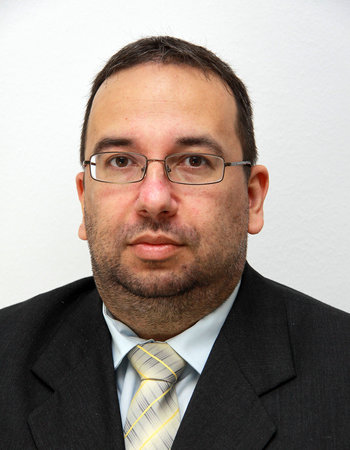
Professor Dr Norbert Kovács
Full professor, deputy clinical director