Relating layer-specific quantitative 7T MRI to histology, gene expression and connectomics
In this project, we elucidate the relationship between layer-specific quantitative Magnetic Resonance Imaging (qMRI) and cytoarchitectonics, gene expression patterns and white matter connectomics in order to provide novel biomarkers and an anatomical precise framework for tracking neurodegenerative diseases.
The human cerebral cortex is composed of distinct cortical layers, which are defined based on cell density, cell size and cell type and are selectively targeted in neurodegeneration. In-vivo high-resolution histology using quantitative MRI (qMRI) at ultra high field strength has the potential to provide cortical layer-specific measures that directly relate to patterns of cell composition. Such measures will enable us to gain mechanistic insights into layer-specific neural processing and pave the way for anatomically precise biomarkers of neurodegenerative diseases like Huntington’s disease that show layer-selective vulnerability. A comprehensive validation of MR-based in vivo histology and characterization of its functional relevance however is still lacking.

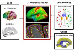
Multi-parametric maps (MPMs) include qMRI parameters of effective transverse relaxation rate (R2*), which is sensitive to both myelin and iron and longitudinal relaxation rate (R1), which is mainly sensitive to myelin and to a lesser extent to iron. As shown in the figure above, we compare cortical layer-specificity for R1 and R2* to the von Economo and Big Brain post-mortem histology atlases and connectomics from diffusion MRI. We also investigate the relationship between 7T MPMs, layer-specific gene expression and Huntington’s disease related genes, using the Allen Human Brain atlas.
We show that R2* across cortical depths is highly correlated with layer-specific cell numbers, cell staining intensity and gene expression. Further, we demonstrate a strong relationship between cortico-striatal and intra-hemispheric white matter connections from high-fidelity diffusion tractography and qMRI across cortical depths.
These findings constitute an important step in the development of UHF qMRI as a tool for in-vivo histology and demonstrate the potential of combining 7T MPMs, gene expression and white matter connections to provide an anatomically precise framework for investigating layer-specific neural processing.


