Quantification of transverse relaxation times of human cortical and subcortical structures in vivo at 7T
An accurate and robust quantification of relaxation parameters T1, T2, and T2* is crucial for studying the microstructural composition of the human brain in vivo. With the developments in ultra-high field (UHF) MRI it has become possible to measure those MR parameters at different cortical depths (Trampel et al., 2019, Neuroimage 197, 707 - 715). However, especially the measurement of the transverse relaxation parameters, at the necessary high spatial resolution, requires either long scan times or sophisticated analysis methods for the distinction of desired from nuisance signals and minimization of measurement bias. Therefore, optimization and analysis of common spin-echo based transverse relaxation measurement techniques towards reducing bias and accurately quantifying transverse relaxation is done.
In a piloting study we compared a number of approaches for quantifying T2 regarding their accuracy, precision, robustness and required scan time. All experiments were conducted at a 7T MRI scanner. Besides a standard spin-echo technique, a multi-echo CPMG (Carr-Purcell-Meiboom-Gill) sequence was applied for determining the intrinsic transverse relaxation T2. For the necessary correction of bias field effects (and resulting stimulated echo signal caused by non-ideal 180 degrees phase reversals), T2 estimation was based on a simulated dictionary of signal curves (Ben‐Eliezer N et al., 2015, Magn Reson Med, 73, 809-817). Additionally, a gradient-echo sampled spin-echo (GESSE) technique was used (Yablonski DA et al, 1997, Magn Reson Med, 37, 872-6) to simultaneously quantify T2 and the effective transverse relaxation time T2*.

T2 maps of a representative slice of the same brain, acquired with different methods with an isotropic resolution of (1 mm)³. Total scan time for each method: SE ~33 min, CPMG ~7 min, GESSE ~6 min, for scanning 8 slices.
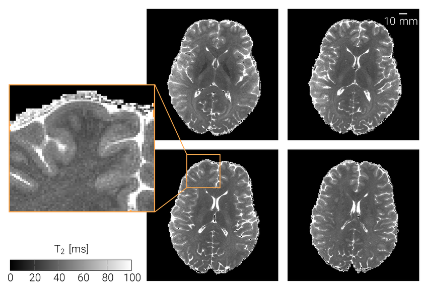
The CPMG method yielded quantitative T2 maps comparable to the SE technique (see Fig 1). Imperfect bias field corrections are still apparent in temporal brain regions, where maintaining B1 field homogeneity at 7T is typically challenging. The GESSE approach also resulted in comparable values (Fig. 1). However, CPMG and GESSE techniques are about 5 times faster than the standard SE method enabling acquisition of sub-millimeter T2 maps (Fig 2). Hence, CPMG and GESSE were identified as potential candidates for quantification of T2 at different cortical depths.

T2 map of 4 brain slices of a human brain acquired with the CPMG sequence and a modelling approach based on a dictionary simulation, the resolution was pushed to (0.6 mm)³ isotropic and the scan time was ~12 min.
It is, however, necessary to fully understand the influence of the MR sequence on the relaxation processes within the tissue of interest under realistic conditions. Currently, the simulation efforts for dictionary creation are expanded to achieve a more accurate description of the expected signal curve and predict the effect of setup bias. Further adaption to incorporate other bias effect like diffusion and motion is planned with the aim of more flexible application for different use cases and other sequences. Furthermore, the single-shot GESSE technique will be used to its full potential enabling the simultaneous measurement of T2 and T2*.


