Laminar fMRI in the human prefrontal cortex at 7T: A replication study of layer-dependent activity during working memory
A recent fMRI study (Finn et al., 2019) demonstrated layer specific responses in the dorsolateral prefrontal cortex (dlPFC) during a working memory task using VASO fMRI at 7T. Superficial layers were more active during the delay period when working memory items needed to be manipulated compared to their mere maintenance. In contrast, deeper layers were more active when participants had to respond by pressing a button. Like many current layer fMRI studies, this study relied on several manual or semi-manual processing steps, including the selection of ROIs. However, to make the leap from methodological demonstration to routine use in cognitive neuroscience research it is important to confirm the robustness of such findings, ideally using a more automatic analysis approach.

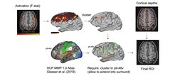
We repeated this experiment in 21 participants and analyzed it using a fully automated and reproducible pre-registered analysis approach. ROIs were determined by combining anatomical information obtained from the HCP MMP 1.0 atlas (Glasser et al., 2016) and on whether a location was activated by the experimental paradigm (Fig.1). We chose the largest activation cluster overlapping p9-46v and selected all contiguous voxels that belonged either to p9-46v or any of the surrounding parcels (46, 8C, IFSp, IFSa) accounting for uncertainties in single subject parcellation. This ROI was then split into a superficial and a deep layer based on cortical depth estimates. Like Finn et al. (2019) we sampled time courses from both layers and analyzed the signal during the delay and response periods.

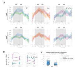
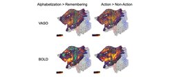
Fig. 2A shows trial averaged VASO time-courses for upper (top row) and deeper layers (bottom row) for alphabetizations vs. remembering (left), action vs. non-action (middle) and differences between conditions from both types of runs. Each panel shows the negative percent VASO signal response, i.e. a proxy of cerebral blood volume change and activation, as a function of trial time. A sequence of 5 random letters were presented at t = 0 s. At t = 4 s a cue indicated whether participants had to alphabetize the sequence or remember it in its original order. At t = 14 s one letter was probed, and participants had to respond using a button press. The shaded band indicates the 95% confidence interval over subjects. Filled circles and open circles represent condition contrasts data points for the delay and response period, respectively. These data points were averaged and further analyzed (Fig. 2B). Our results do not show the same layer specific effects as Finn et al. (2019). The interaction between type of contrast and layer was not significant for either period. There is a non-significant trend for higher superficial layer delay period activity that is specific to manipulation. Also, bootstrapping of interaction effect sizes (Fig. 2C) suggest that a potential small effect, which we may not be sufficiently sensitive to, is expected to be much smaller than reported by Finn et al. (2019). Importantly we did not find any evidence for a deep layer effect for motor responses. Furthermore, an exploratory group analysis of experimental contrasts resulted in task specific activation in an extensive set of lateral prefrontal and parietal regions beyond our ROI (z-stats on flattened cortex representation shown in Fig. 3) with similar activation time courses and layer profiles during this task.

We conclude that evidence about the type of laminar activity in the human dlPFC during working memory tasks is still inconclusive. The wide distribution of involved regions underlines the need for a principled and reproducible definition of ROIs. It is necessary to further investigate which methodological differences may have caused differences in results between studies.


