Mapping ocular dominance columns in humans using GE-EPI, SE-EPI and SS-SI VASO at 7 T
With the advent of functional magnetic resonance imaging (fMRI) at ultra-high magnetic fields (≥ 7 T), mapping of small physiological structures like ocular dominance columns (ODCs) in the primary visual cortex (V1) become feasible in vivo (Yacoub et al., 2007, Neuroimage 37, 4, 1161–77). However, gradient-echo based sequences (GE-BOLD), which are the usual workhorse for fMRI, are inherently sensitive to large draining veins (Polimeni et al., 2010, Neuroimage, 52, 4, 1334–46), which degrade their specificity towards the source of neural activation. Other approaches like spin-echo based (SE-BOLD) (Chaimow et a., 2018, Neuroimage, 164, 32–47) and cerebral blood-volume weighted sequences (SS-SI-VASO) (Huber et al., 2017, Neuron, 96, 6, 1253–63) promise higher specificity at the expense of sensitivity.
In this study, we compared these different approaches in their sensitivity to functionally detect ODCs in several humans. Four participants were scanned at 7T in several sessions. In functional mapping sessions, ODCs were localized by alternate visual stimulation of single eyes using moving sparse random dot stereograms viewed through in-house constructed anaglyph goggles (Nasr et al., 2016, J Neurosci, 36, 6, 1841–57). All functional data were acquired with a nominal isotropic voxel size of (0.8 mm)3.
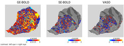
Figure 1 shows thresholded z-score maps of ODCs (left eye > right eye) for GE-BOLD (left), SE-BOLD (middle), and SS-SI VASO (right). Data are illustrated on a flattened cortical surface mesh, which only represents the stimulated region in V1. The expected topography of alternating left-eye and right-eye columns can be seen in all plots. Black dots are added as a reference to aid comparison between maps. The dashed white line outlines the region, which was outside of the imaging volume for SE-BOLD and VASO measurements. As expected, GE-BOLD showed the largest response, while responses for VASO were barely detectable. Note that VASO shows the inverse contrast due to signal decrease associated with increases in CBV.

Our results demonstrate the possibility of measuring ODCs in humans using GE-BOLD, SE-BOLD, and SS-SI-VASO in terms of sensitivity. To the best of our knowledge, this study shows for the first time ODCs in humans with SS-SI-VASO.


