A multi-modal approach to in vivo U-fibre mapping in the early visual processing stream
Short cortico-cortical association fibres (U-fibres) are white matter fibres that run directly below the cortical grey matter in the superficial white matter and connect nearby cortical areas. U-fibres have demonstrated involvement in brain development, function and pathology but are underrepresented in the current human brain connectome. A more complete picture of the human brain connectome can be obtained by reliably mapping the U-fibres, but this requires high quality sub-millimetre resolution in vivo diffusion MRI, dedicated fibre and tractography models and appropriate validation.
We addressed U-fibre connectivity mapping by acquiring sub-millimetre resolution in vivo diffusion MRI facilitated by the high performance gradients (300 mT/m maximum gradient amplitude) of the 3T Connectom scanner (Siemens Healthineers, Erlangen, Germany) and targeted validation via mapping U-fibre connectivity in the human brain between the primary and secondary visual cortical areas (V1 and V2, respectively) which are known to be retinotopically organised. The detected U-fibre connectivity maps were found to be retinotopically organised, i.e., connections between corresponding retinotopic areas of V1 and V2 were relatively higher.
This proof-of-concept study showcases robust U-fibre connectivity mapping in vivo. We believe that the current research effort — combining multiple MRI modalities for U-fibre mapping and validation — is an essential step toward the construction of a more complete human brain connectome.
Related publications:
Movahedian Attar F, Kirilina E, Haenelt D, Trampel R, Edwards L J, Weiskopf N. 2020. Mapping Short Association Fibers in the Early Cortical Visual Processing Stream Using In Vivo Diffusion Tractography. Cerebral Cortex. doi: 10.1093/cercor/bhaa049
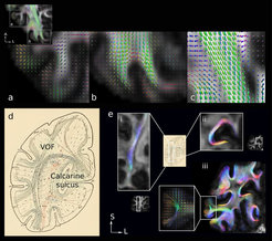
 estimates and tractography were enabled at sub-millimitre resolution. (a) intra-cortical radial fibres, (b) sub-cortical superficial white matter fibres and (c) fibres in the optic radiation tract were reconstructed. (d) histological map of short white matter fibre connections in the occipital lobe. (e) tractography reconstructed the vertical occipital fasciculus (VOF), fibres connecting the upper and lower calcarine sulcus and other U-shaped fibres in the histology map in vivo.")