In Parkinson's disease, the depletion of iron-rich dopaminergic neurons in nigrosome 1 in substantia nigra precedes first motor symptoms by almost two decades. Methods capable of monitoring this neuronal depletion at an early disease stage are highly desired for diagnosis and treatment monitoring.
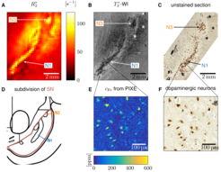
Fig 1: Quantitative histology and MRI on a representative substantia nigra specimen. A: On a quantitative R2* map of substantia nigra, nigrosomes nigrosome 1 and 3 (N1 and N3) are visible as hyperintense areas. B: On 50 µm resolution T2*-weighted images of substantia nigra, granular hypointensities resembling dopaminergic neurons are visible in nigrosome 1 and 3. C: An unstained tissue section including substantia nigra shows nigrosome 1 and 3 as dopaminergic neurons-rich areas (dopaminergic neurons enlarged for better visibility). D: Subdivision of Substantia Nigra (SN; Damier et al., 1999, Brain, 122, 8, 1437–1448) shows elongated nigrosome 1 and circular nigrosome 3. E: On a quantitative iron map of nigrosome 1 obtained with PIXE, neuromelanin domains within dopaminergic neurons show increased iron concentration. F: On light microscopy images of the same area as in (E), the brown neuromelanin-pigmented part of dopaminergic neurons is visible.
Fig 1: Quantitative histology and MRI on a representative substantia nigra specimen. A: On a quantitative R2* map of substantia nigra, nigrosomes nigrosome 1 and 3 (N1 and N3) are visible as hyperintense areas. B: On 50 µm resolution T2*-weighted images of substantia nigra, granular hypointensities resembling dopaminergic neurons are visible in nigrosome 1 and 3. C: An unstained tissue section including substantia nigra shows nigrosome 1 and 3 as dopaminergic neurons-rich areas (dopaminergic neurons enlarged for better visibility). D: Subdivision of Substantia Nigra (SN; Damier et al., 1999, Brain, 122, 8, 1437–1448) shows elongated nigrosome 1 and circular nigrosome 3. E: On a quantitative iron map of nigrosome 1 obtained with PIXE, neuromelanin domains within dopaminergic neurons show increased iron concentration. F: On light microscopy images of the same area as in (E), the brown neuromelanin-pigmented part of dopaminergic neurons is visible.
MRI is particularly suited for this task, since it is sensitive to iron accumulated in the neuromelanin of dopaminergic neurons (Fig. 1). However, the mechanisms of MRI contrast in substantia nigra are unknown, hindering the development of specific biomarkers. We elucidate the mechanisms of iron-induced transverse relaxation in nigrosome 1 by combining quantitative 3D iron histology, quantitative MRI on post mortem human brain tissue, and biophyiscal modeling.
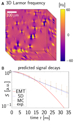
Fig 2: Biophysical modeling of gradient echo signal in nigrosome 1. A: Dopaminergic neurons were strong Larmor frequency perturbers on frequency maps predicted from quantitative iron histology. B: Gradient echo signal predicted in Static Dephasing regime (SD; Yablonskiy and Haacke, 1994, MRM, 32, 6, 749-63) agree much better with the experimental signal decay and Monte Carlo simulations (MC; Gagnon et al., 2015, J. Neurosci., 35, 8, 3663–3675) than predictions from Effective Medium Theory (EMT; Kiselev and Novikov, 2002, PRL, 89, 27, 278101). The static dephasing regime allows for linking the iron-induced R2* to the total iron content in dopaminergic neurons.
Fig 2: Biophysical modeling of gradient echo signal in nigrosome 1. A: Dopaminergic neurons were strong Larmor frequency perturbers on frequency maps predicted from quantitative iron histology. B: Gradient echo signal predicted in Static Dephasing regime (SD; Yablonskiy and Haacke, 1994, MRM, 32, 6, 749-63) agree much better with the experimental signal decay and Monte Carlo simulations (MC; Gagnon et al., 2015, J. Neurosci., 35, 8, 3663–3675) than predictions from Effective Medium Theory (EMT; Kiselev and Novikov, 2002, PRL, 89, 27, 278101). The static dephasing regime allows for linking the iron-induced R2* to the total iron content in dopaminergic neurons.
We developed a comprehensive biophysical model accounting for the chemical form of iron binding and the heterogeneous iron distribution at the cellular scale. This model was informed with 3D quantitative iron concentration maps of nigrosome 1 obtained from combining Proton-Induced X-ray Emission microscopy (PIXE) with iron histochemistry. We showed that iron in dopaminergic neuron is the dominant source of effective transverse relaxation rate R2*. We determined the proper theoretical relaxation regime describing R2*, which was found to be close to static dephasing (Fig. 2). In this regime, R2* is analytically linked to the total iron content in dopaminergic neurons, i. e., the product of neuronal density and mean cellular iron concentration (Yablonskiy and Haacke, MRM, 1994, 32, 6, 749-63). Our model’s predictions were shown to be accurate by comparing them to relaxation rates acquired at 7 T on a specimen before and after chemical iron extraction.
For the first time, we achieved a mechanistic model of iron-induced MR contrast in substantia nigra derived from first principles and based on iron microstructure quantification. This knowledge paves the road toward novel, specific biomarkers for Parkinson's disease.
We investigate the relationship between quantitative MRI (qMRI) at different cortical depths and cell counts, gene expression and white matter connections in the brain in order to provide novel biomarkers for tracking neurodegenerative diseases.
Robust U-fibre connectivity mapping can be achieved in vivo in the early visual processing stream using combined diffusion weighted imaging and functional retinotopy
In this project, we investigate the brains of wild chimpanzees who died of natural causes at different developmental stages using high-resolution quantitative MRI and histology.
Understanding brain development and decline is of utmost importance in an aging society. MRI Biophysics Research Group aims to uncover crucial mechanisms of human brain aging, by identifying the contribution of iron accumulation, a major determinant of brain development and brain decline.
We characterize the cortical layers by biomechanical modeling and simulation of the developed human cortex tissue in-vivo using hyperelastic material models.
We used high-resolution fMRI and multivariate pattern analysis (MVPA) to explore how attentional modulation of working memory affects laminar specific representations in dorsolateral prefrontal cortex (dlPFC).
We performed laminar fMRI during a delayed match-to-sample task and varied working memory load and the requirement for a motor response. We found layer specific univariate and multivariate effects.
A recent fMRI study showed layer-specific responses in the dorsolateral prefrontal cortex during a working memory task. We attempted to replicate the original findings using newly acquired data and a fully automated analysis.
Using a field strength of 7 Tesla, the "Arterial Blood Contrast" (ABC), which is based on the Magnetization Transfer effect, could be measured with an isotropic spatial resolution of 1.5 mm in combination with a conventional functional MRI contrast.
In this project, we studied cortical myelin in living humans at the spatial scale of cortical columns using high-resolution quantitative magnetic resonance imaging (MRI) methods at 7 T.
 are visible as hyperintense areas. B: On 50 µm resolution T2*-weighted images of substantia nigra, granular hypointensities resembling dopaminergic neurons are visible in nigrosome 1 and 3. C: An unstained tissue section including substantia nigra shows nigrosome 1 and 3 as dopaminergic neurons-rich areas (dopaminergic neurons enlarged for better visibility). D: Subdivision of Substantia Nigra (SN; Damier et al., 1999, Brain, 122, 8, 1437–1448) shows elongated nigrosome 1 and circular nigrosome 3. E: On a quantitative iron map of nigrosome 1 obtained with PIXE, neuromelanin domains within dopaminergic neurons show increased iron concentration. F: On light microscopy images of the same area as in (E), the brown neuromelanin-pigmented part of dopaminergic neurons is visible.")
 agree much better with the experimental signal decay and Monte Carlo simulations (MC; Gagnon et al., 2015, J. Neurosci., 35, 8, 3663–3675) than predictions from Effective Medium Theory (EMT; Kiselev and Novikov, 2002, PRL, 89, 27, 278101). The static dephasing regime allows for linking the iron-induced R2* to the total iron content in dopaminergic neurons.")