Cortical myelination in chimpanzees
Comparing brain ontogeny across hominoid species provides important insights into the evolution of human cognition and behaviour. However, developmental studies on cortical brain maturation in great apes are rare and mostly rely on captive primates. The captive environment may not fully promote brain plasticity, and primates raised in captivity do not express their typical entire behavioural repertoire. In this unique, collaborative project, we study cortical myelination in whole post-mortem brains of wild and captive chimpanzees and other great apes at different developmental stages, by combining ultra-high resolution quantitative magnetic resonance imaging with histology.
In the pilot phase of the project, we studied the brains of six chimpanzees who died from natural causes (between 3 weeks and 32 years old). The tissue was collected and formalin-fixed in pH 7.4 buffered solution within hours after death. The high tissue quality enabled myelin-sensitive MRI multi-parameter mapping at high field strength with 300 μm isotropic resolution. The combination of whole-brain coverage and ultra-high resolution allowed characterization of myeloarchitecture across the entire brain. Whole-brain MRI-measures were validated by various histological methods in selected brain regions.
We have established MRI-based mapping of myeloarchitecture in great apes at unprecedented resolution, providing a unique resource for comparative neuroscience research. Combined with white matter connectomics and behavioural characterisation of the same individuals (conducted by our collaborators at the Dept. Neuropsychology and MPI-EVA), these data will open new doors for a better understanding of the functional neuroanatomy underlying human-specific traits.

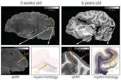
The figure shows qMRI maps of two wild chimpanzees at different developmental stages. Various quantitative MR parameters (R1, R2*, PD and MT saturation) were acquired in a human 7 Tesla MRI scanner (Siemens Healthineers, Erlangen, Germany) with ultra-high 300μm isotropic resolution. Sagittal slices of the longitudinal relaxation rate (R1) are shown for two chimpanzees – a 3 week old and a 6 year old. Early myelination (EM) in white matter in the chimpanzee newborn is clearly visible in qMRI (R1) and the myelin histology stain (anti-myelin basic protein antibody; right column). In the 6 year old chimpanzee, the myeloarchitectonically distinct primary and secondary visual areas (V1 and V2) are clearly visible in qMRI (R2*) and the myelin histology stain (anti-myelin basic protein antibody; right column).


